ELMT 101: Calming skincare you can trust (with science to prove!)
by Gina Myung on Jan 13, 2022

What is a wound healing assay?
A wound-healing assay is a simple, and one of the earliest developed methods to study cell migration direction in vitro*, which mimics cell migration during wound healing in vivo**.- ● in a test tube, culture dish, or elsewhere outside a living organism*
- ● in a person, or a living organism**
Why the wound healing assay?
The reason this method of testing was chosen is because, as you probably know, the Advanced Calming Solution’s main function is to calm the skin. When skin is damaged, it is usually followed by irritation like redness, inflammation pain and fever, especially in the case the wound can not recover properly. Therefore, if the use of the calming solution can ensure significant recovery of skin damage, we can prove that it effectively helps calm the skin.
elmt
Advanced Calming Solution
How do we prepare?
Before we start, because the study is conducted in vitro, the cells are more exposed and vulnerable as compared to when they’re in the form of living cell tissue in our body. So as to ensure the viability of the cells, the study is conducted with diluted amounts of the solution. In order to determine which percentages to conduct this at, a cytotoxicity test is performed.
For this process, the same type of cells that were going to be used in the wound healing assay, which in this case were human dermal fibroblast cells, were thawed and grown for 24 hours. Then, different concentrations of the Advanced Calming Solution were added and the plates were incubated for 24 hours.
Once researchers were able to find the correct concentrations, 0.125% and 0.25%, after analyzing the 24 hour results, the wound healing assay was conducted.

What are the steps?
The study was conducted using Human Dermal Fibroblast cells, the most common type of cell found in connective tissue. Fibroblasts secrete collagen proteins that are used to maintain a structural framework for many tissues and also play a key role in wound healing.
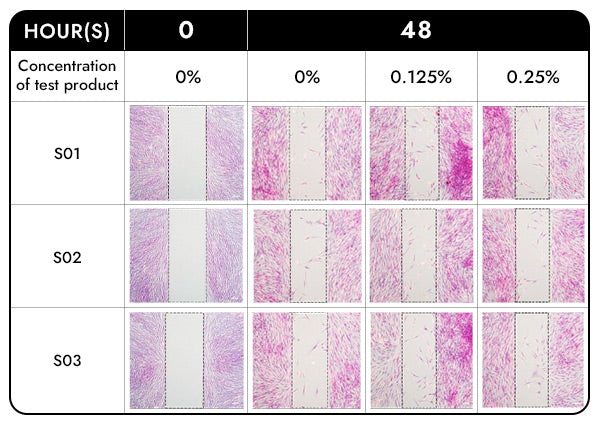
Cell migration was assessed using a cell culture monolayer, consisting of two wells that were separated by a culture insert that acted as a wall. The culture inserts were removed to form a gap between the cells, which were then washed to remove cell debris and cultured with different concentrations of the calming solution and incubated 48 hours.
Cells were stained purple with a staining solution so they could be photographed and analyzed using a cell imaging program. Photographs were taken at 0 hours (as a control), and after 48 hours.

Conclusion
In comparison to the control group, the wound recovery rate (%) was found to be 470.516% (approximately 4.7 times) at 0.125% concentration of the test product and 460.254% (approximately 4.6 times) at 0.25% concentration of the test product in significantly increasing the migration of human dermal fibroblasts.


